Scanning Near-Field Microscopy (SNOM)
Application Description
Scanning near-field microscopy (SNOM) is a type of scanning probe microscopy technique that measures light-matter interactions at the nanoscale to investigate local optical properties of a sample. It relies on optical near-field effects to push its resolution below the diffraction limit as obtained in conventional far-field measurements. SNOM techniques first used a tapered optical fiber to create an optical aperture, but recent years saw the development of so-called apertureless SNOM or scattering SNOM, where the incident light is scattered by a metal-coated AFM probe.
In this way, nano-optical cavities can be formed to enhance and control the confined light by modifying the local electromagnetic environments underneath the tip. SNOM is thus used in various fields such as tip-enhanced Raman spectroscopy (TERS) and THz spectroscopy to study plasmonic devices, and more generally in nano-optical experiments.
Measurement Strategies
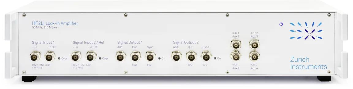
Incident light shed on a metallic AFM tip is scattered at the modulation frequency of the cantilever's mechanical motion. Non-linear optical processes arise in the nano-cavity formed by the AFM tip and the sample surface: the reflected light thus carries information about the sample's optical properties to the optical detector. The detection of all harmonic and sideband signals can be achieved thanks to multi-frequency lock-in amplifiers such as the Zurich Instruments HF2LI or UHFLI, depending on the requirements of the experiment in terms of optical modulation scheme and demodulation bandwidth. As there exist many different SNOM configurations, the figure illustrates in a broad context the optical and mechanical mixing that happens under the tip and that is detected by an HF2LI Lock-in Amplifier through multiple inputs and demodulators.
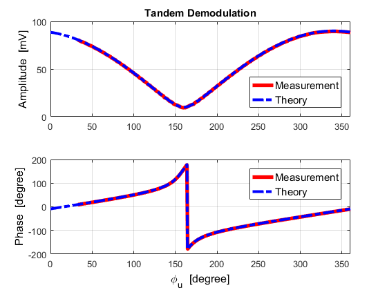
The insert in the figure corresponds to the detected response of frequency components that arise from the modulation sources, typcially between a carrier or cantilever at fc and a modulation at fm. The signal from the optical detector is captured at the lock-in input and demodulated in 2 stages (also called tandem demodulation): a wide-band demodulator measures frequency components around a harmonic of the carrier, the output of which is then fed back into the second lock-in input to demodulate each sideband separately with a narrow band. This makes it possible to recover and image the optical phase and amplitude of each sideband from the scattered light as the tip scans the surface. To benefit from maximum signal enhancement at the resonance frequency of the mechanical sensor, a phase-locked loop (PLL) and automatic gain controller can be introduced in the mechanical path for resonance tracking.

Product Highlights

The Benefits of Choosing Zurich Instruments
- Simplify your setup: perfect synchronization is ensured between optical and mechanical modulation as the same instrument controls both signal generation and detection.
- You can extend the amount of information you acquire by performing multiple higher-harmonic-generation measurements at once thanks to a broadband multi-demodulation scheme at up to 5 MHz within a 600 MHz input bandwidth.
- Don't miss any sidebands required to understand the complete non-linear optical response of the sample. A higher number of demodulators (> 8) can be accessed with multi-device synchronization (MDS), enabling your setup to grow with your needs.
- The time- and frequency-domain analysis toolset of the LabOne® user interface allows you to better understand and optimize the rich and complex frequency mixing arising from electro-optical interactions at the tip apex.